3D Breast Ultrasound
About 40 % of women have dense breast tissue.
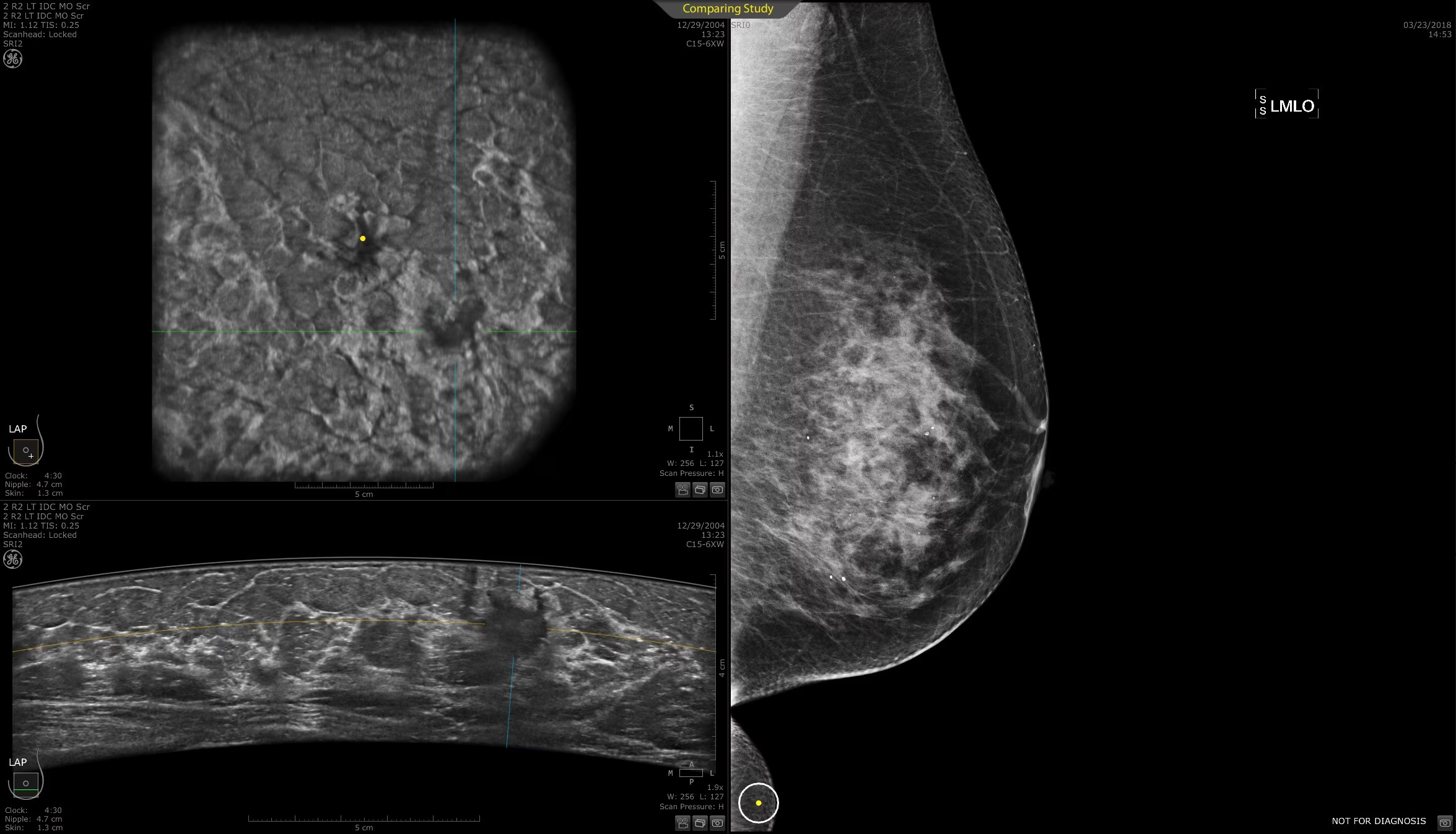
Having dense breasts is normal—it means your breasts contain more tissue than fat. For these women, mammograms alone may not be enough to detect breast cancer. On a mammogram, dense tissue and masses both appear white, so a suspicious lump may be hidden in the dense tissue. When dense tissue is scanned with ultrasound, tissue appears white and masses appear black, making them easier to see.
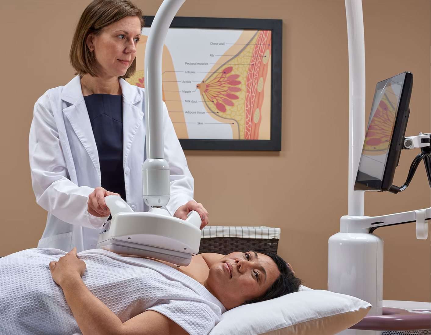
ABUS captures multiple angles of the breast, including coronal (top-down), sagittal (side) and transverse (front-to-back) planes. Each scan produces hundreds of high-resolution images that the system compiles into a 3-D model of the breast. Radiologists can scroll through these layers to examine tissue at various depths, improving their ability to detect abnormalities that might be missed in 2-D images.
Invenia ABUS 2.0 is the only ultrasound technology FDA-approved for breast-cancer detection in women, specifically developed to help doctors find cancers hidden in dense breast tissue that may be missed by mammography. Invenia ABUS screening is completely unlike a mammogram: a layer of lotion is applied to your breast, and a scanner acquires images in about 15 minutes—without heavy compression or radiation.
Advantages of ABUS (Automated Breast Ultrasound System)
- Women with dense breast tissue, breast implants and younger women benefit significantly from ABUS.
- Dense tissue can obscure abnormalities on mammograms because both tissue and tumors appear white; ABUS uses sound waves to create clearer imaging.
- In women with implants, traditional mammography can hide parts of the breast; ABUS improves visibility.
- Younger women who may wish to avoid radiation exposure can use ABUS as a safe, effective screening method.
About Us
Our sonographers have 20 years of diagnostic experience and are licensed by the American Registry for Diagnostic Medical Sonographers (ARDMS). All procedures are interpreted by U.S. board-certified radiologists specialized in ABUS readings.
- What it shows: Subtle lesions, cysts or masses that may be hidden on mammography.
- Procedure length: 15–25 minutes per breast.
- Why choose it: Non-invasive, radiation-free supplemental screening for women with dense tissue or inconclusive mammograms.
- Your experience: You lie face-up as the automated transducer sweeps over each breast; minimal compression ensures comfort.
- Results: High-resolution 3D datasets interpreted by board-certified radiologists; report delivered within 24 hours.
- Insurance & payment: We accept Medicare, PPOs, HMOs and cash; no prior authorization required for HMO plans.
