Echocardiogram
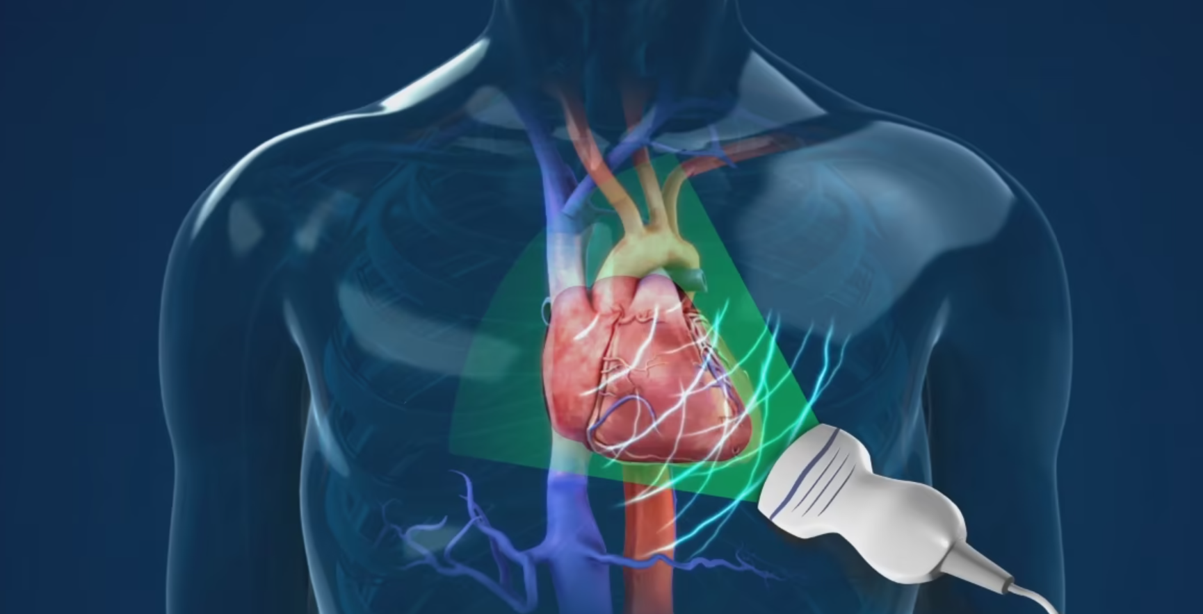
An echocardiography, echocardiogram, cardiac echo, or simply an echo, is an ultrasound of the heart. It is a type of medical imaging of the heart, using standard ultrasound or Doppler ultrasound.
Echocardiography has become routinely used in the diagnosis, management, and follow-up of patients with any suspected or known heart diseases. It is one of the most widely used diagnostic imaging modalities in cardiology. It can provide a wealth of helpful information, including the size and shape of the heart (internal chamber size quantification), pumping capacity, location and extent of any tissue damage, and assessment of valves. An echocardiogram can also give physicians other estimates of heart function, such as a calculation of the cardiac output, ejection fraction, and diastolic function (how well the heart relaxes).
Echocardiography is an important tool in assessing wall-motion abnormality in patients with suspected cardiac disease. It helps in reaching an early diagnosis of myocardial infarction by showing regional wall-motion abnormality. It is also important in treatment and follow-up in patients with heart failure, by assessing ejection fraction.
- What it shows: Heart chamber size, valve motion, ejection fraction, blood flow patterns and congenital anomalies.
- Procedure length: 30–45 minutes.
- Why choose it: Radiation-free, detailed functional assessment, vital for diagnosing valve disease, heart failure, cardiomyopathy and more.
- Your experience: You lie on your left side as the sonographer places the transducer on various chest locations; you may be asked to hold your breath briefly.
- Results: Comprehensive report from our cardiology team delivered within 24 hours.
- Insurance & payment: We accept Medicare, PPOs, HMOs and cash; no prior authorization required for HMO plans.
